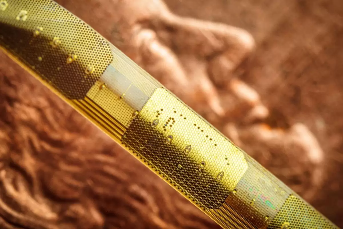
Researchers use wireless sensor 5 times thinner than hair to map brain
Share- Nishadil
- January 17, 2024
- 0 Comments
- 4 minutes read
- 298 Views

Researchers use wireless sensor 5 times thinner than hair to map brain
UC San Diego researchers unveil a revolutionary brain monitoring system, enabling high resolution, wireless recording in deep brain structures for diverse clinical applications.
The Integrated Electronics and Biointerfaces Laboratory (IEBL) at the University of California San Diego has introduced a revolutionary approach to clinically recording deep brain activity. The research unveils a novel sensor manufacturing technique that enables minimally invasive, high resolution recording as deep as 4 inches (10 cm) inside the human brain.
Innovative sensor manufacturing approach The unique approach hinges on ultra thin, flexible, customizable probes constructed with clinical grade materials. These probes are equipped with sensors capable of recording extremely localized brain signals. Notably, their smaller size allows for close placement, enabling high resolution sensing in specific areas at unprecedented depths within the brain.
electrical engineering professor Shadi Dayeh, the corresponding author on the paper, explains, “We developed an entirely different manufacturing method for thin film electrodes that can reach deep brain structures at a depth that is necessary for therapeutic reasons enabling reproducible, customizable, and high throughput production of electrodes but with a high spatial resolution and channel count despite a thinner electrode body.” The probes, only 15 microns thick (about 1/5th the thickness of a human hair), can currently record with up to 128 channels, a significant leap compared to today’s clinical probes with only 8 to 16 channels.
The researchers envision further expansion, with thousands of channels per probe potential. This advancement promises to dramatically enhance physicians’ ability to acquire, analyze, and understand brain signals at a finer resolution. The implications of this technology extend beyond , with potential applications in Parkinson’s disease, movement disorders, obsessive compulsive disorder, obesity, treatment resistant depression, high impact chronic pain, and various other disorders.
The UC San Diego Micro stereo electro encephalography (µSEEG) is a crucial step toward wireless monitoring of patients with treatment resistant epilepsy for extended periods, up to 30 days. Professor Dayeh envisions a wireless system allowing patients to move freely within the hospital environment and at home while continuously monitoring cortical and deep brain structures.
Wireless brain monitoring system on the horizon The researchers are actively working toward their vision of a brain monitoring system with sensors inserted deep within the brain and on the surface. This system, communicating wirelessly with a small computer system in a cap, could provide and computational infrastructure for recording brain signals for up to 30 days.
Dr. Dayeh emphasizes, “The ultimate goal is to advance the system and related required technologies by 2026 to give patients access to a wireless system that allows them to move freely.” The study reports the successful functioning of the system in two human patients during tumor removal surgeries.
Data from various animal models, including rats, pigs, and non human primates, demonstrate the system's versatility. Up to 10 cm in length, the probes allowed recording from deep brain structures, providing crucial insights into brain dynamics at macroscopic and single neuron levels. Evaluating the impact: Insights from authors Dr.
Keundong Lee, the first author, expresses satisfaction with the innovative manufacturing technique, emphasizing its potential for high resolution and minimally invasive diagnosis. Dr. Angelique Paulk notes the significant progress from testing in 2018 to a version much closer to clinical use. Dr. Yun Goo Ro highlights the excitement of seeing his Ph.D.
research translated from the benchtop to the bedside, maximizing clinical impact. The new electrode technology holds great promise for reshaping clinical practice, particularly in epilepsy treatment. Dr. Sharona Ben Haim envisions the potential to send patients home during intracranial evaluation, providing longer recording periods and more robust information for precision treatment.
Dr. Eric Halgren emphasizes the transformative potential of discerning individual voices in brain activity, akin to learning the basic vocabulary of thought. Dr. Ahmed Raslan underscores the uniqueness of the depth electrodes, combining higher resolution recording with stimulation capability. The wireless connectivity opens doors to unrestricted environment recording, allowing sampling of various behaviors.
Dr. Sydney Cash sees the potential to understand both normal brain function and pathology better, offering new ways to help people suffering from epilepsy and neurological problems. In conclusion, the pioneering work of the UC San Diego researchers marks a significant stride in brain monitoring, promising transformative applications in clinical settings.
As the team continues refining and advancing this technology, the future of the holds the prospect of wireless brain monitoring systems ushering in a new era of precision medicine for neurological disorders. The research was published in the January 17, 2024, issue of Nature Communications..
Disclaimer: This article was generated in part using artificial intelligence and may contain errors or omissions. The content is provided for informational purposes only and does not constitute professional advice. We makes no representations or warranties regarding its accuracy, completeness, or reliability. Readers are advised to verify the information independently before relying on


